The SOPRO Camera: A revolutionary instrument in cavity detection
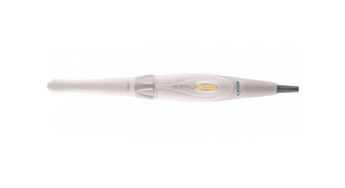
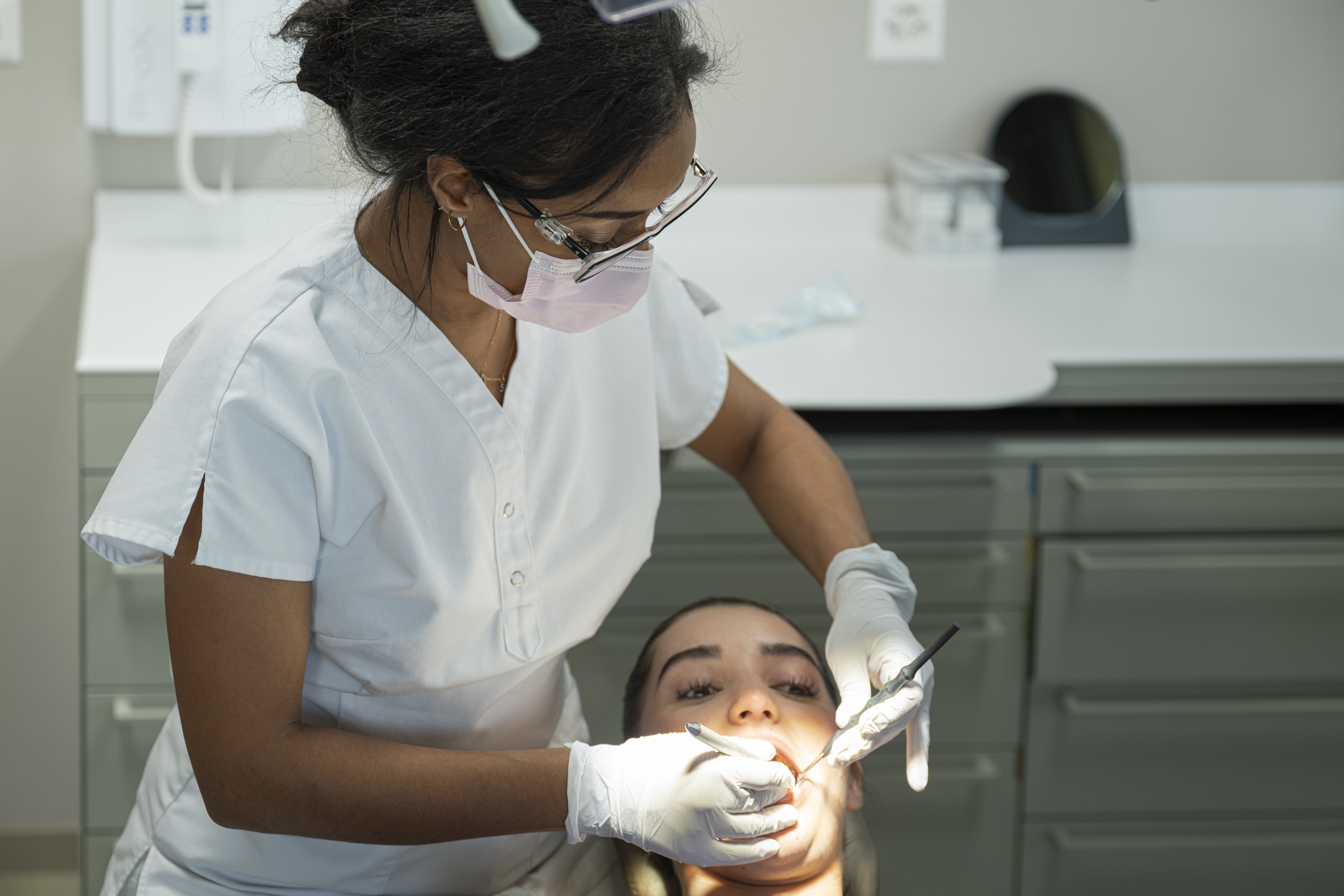
The Smile and Care dental practices in Eaux-Vives and Grand-Saconnex are now equipped with the revolutionary SOPRO intraoral camera. Now a global standard for high-resolution imaging, this camera opens up new perspectives for cavity diagnosis and periodontal treatments. Today, medical imaging has made a significant contribution to improving diagnoses and the widespread use of less invasive interventions.
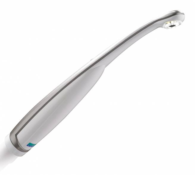
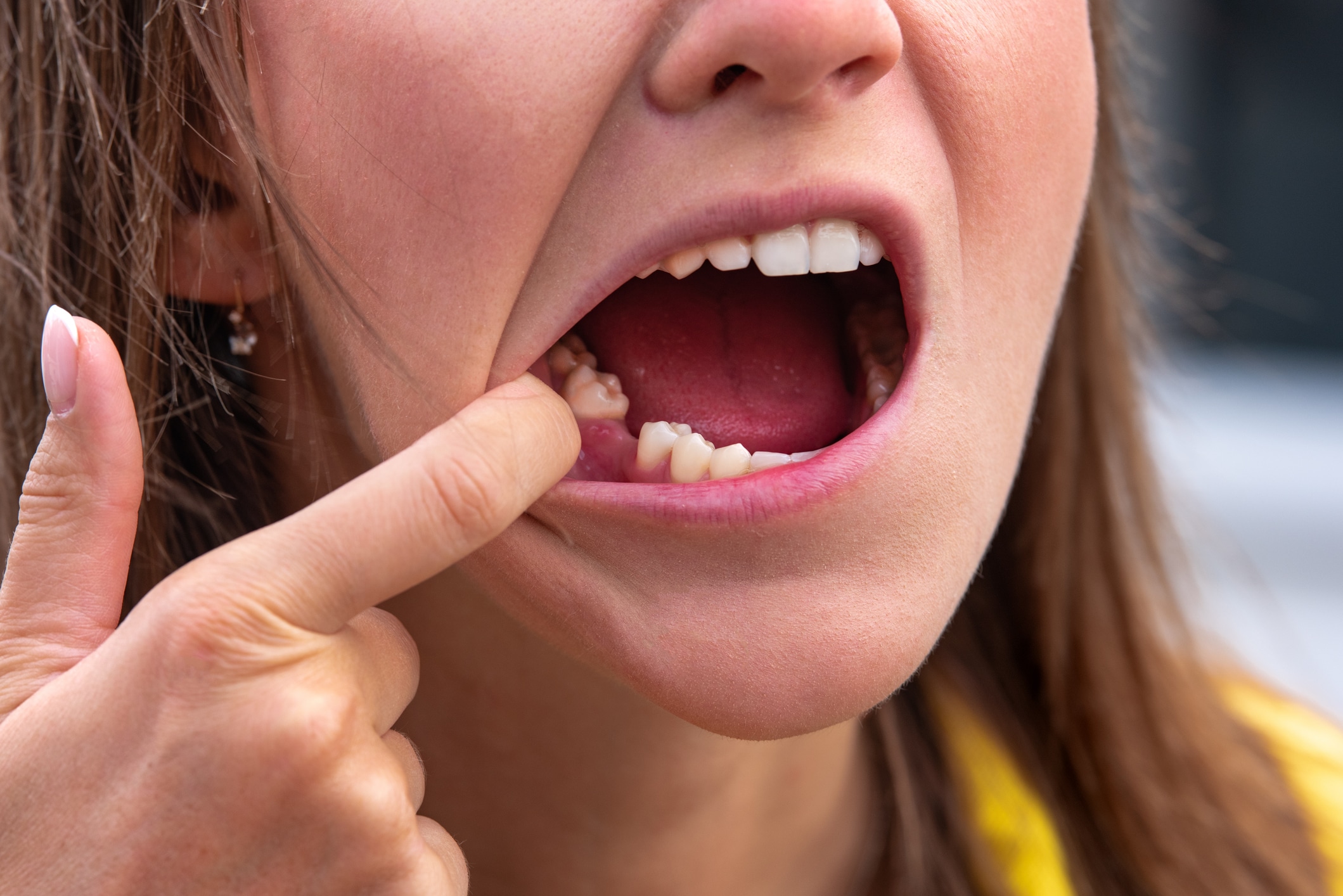
The camera instantly highlights cavities, plaque, and gum inflammation.
This new instrument allows us to carry out a complete and rapid assessment of your oral health. The camera has the SOPRO patent based on fluorescence.
The benefits of autofluorescence
The camera lights up any dental tissues with the necessary wavelength. The material examined absorbs the energy and returns it as fluorescent light. This technique allows us to superimpose the resulting image on the natural anatomical image, thereby revealing the nuances of each tissue that is invisible to natural light. In CARIO mode, it reveals cavities, while in PERIO mode, shows both new and old plaque. The camera is also the first that can reveal gum inflammation in relation to plaque. Thanks to the wavelength emitted by the camera, we are able to highlight the different tissues shown through chromatic mapping.

Dental lesions are shown using a simple colour panel. Gum inflammation appears in a range from pink to purple. New plaque is shown in grainy white, while old plaque appears as an orange-yellow colour.
Numerous patient benefits
This cutting-edge instrument offers numerous patient benefits. Among other things, it enables:
- Make more accurate diagnoses for the assessment of carious lesions

- Offer Improved vision during clinical examination
- Reveal cavities earlier, thus giving our patients the option of less invasive treatment and ensuring the preservation of dental structure.

This tool enables the detection of advanced pathologies and nascent abnormalities thanks to upstream and minimally-invasive intervention.
- Protect our patients by limiting radiological exposure. Indeed, fluorescence imaging pushes the boundaries of digital radiology in the detection of lesions in the hard tissue. This imaging technique ensures our patients suffer no deleterious effects.
- Eliminate uncertainties, as fluorescence facilitates treatment while improving its efficiency and accuracy. It enables the easier differentiation between healthy and infected tissues, and determines carious cavities and their appropriate treatment. Dental pulp is therefore preserved.
The pulp is located in the endodontic part of the tooth (its most internal part). It plays a crucial role in ensuring the formation of dentin – the tooth’s supporting tissues.
- Communicate with our patients with the greatest transparency: Good imagery is worth far more than a long speech, improves understanding of the process, and ensures optimal final results.

The imagery captured is shared with the patient.
The advantages of this state-of-the-art camera
- The intraoral camera offers a 105° view for improved exploration of distal areas. Its rounded shape and the thinness of the distal part make it more comfortable in the mouth.
- Its aspheric lens avoids distortion and provides a high-quality image.

As you can see, the SOPRO camera has become a global standard for dentists seeking high-quality imaging.
At Smile and Care, our quest for innovation is ongoing. We are proud to be able to offer this cutting-edge technology to our patients.

Care and emergencies 6 days a week, Monday to Saturday
